Genetics
Treatment up to Age 56 years old
100% Anonymous Donor Options
EU Standard Care (ISO Certified)
Immediate Start (No Waiting Lists)
Save ~60% vs. UK/EU Prices
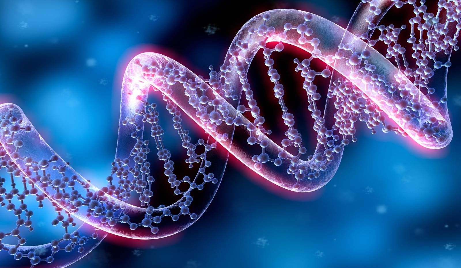
Genetic testing is a way to examine a person’s DNA to check for unusual genes or inherited health conditions. One method involves studying a single cell (called a blastomere) taken from an embryo created through in vitro fertilization (IVF), where one sperm is used to fertilize an egg. This helps doctors understand the embryo’s genetic makeup.
To help patients with recurring failed IVF cycles, genetic diseases, or recurrent miscarriages conceive, we offer PGD, NGS, and other types of chromosomal and genetic test choices based on the patient’s medical history. Our patients give birth to healthy infants because these tests allow our specialists to transfer the chosen, genetically healthy embryos and gather accurate information about the disease. We are aware that IVF is inspired by evolving genetic testing tools. We are convinced that the genetic testing opportunities available in our clinic will benefit our patients.
Because it can help identify genetic problems that children may inherit, clinical genetics is important for IVF. For instance, children may inherit a hereditary condition if one or both potential parents have a genetic mutation that causes it. This can increase the likelihood of a successful pregnancy and a healthy kid by helping to identify embryos free of genetic abnormalities prior to implantation.
Genetic testing will make it possible to identify the presence of hereditary diseases or characteristics, such as Huntington’s disease, sickle cell disease, and cystic fibrosis illness. The decision about whether or not to implant embryos is made after these problems are discovered at a very young age, lowering the likelihood that your possible child may have heritable genetic illnesses.
We can detect chromosomal abnormalities that may affect the success of the IVF procedure and the potential life of the kid by using genetic testing to screen for chromosomal stability in your embryos. While the most promising embryo can be implanted, it is also highly useful because it is likely to be linked to a successful pregnancy and a healthy child.
Results could be improved when combined with the addition of genetic testing to the IVF procedure. By having access to your embryos’ genetic profile, we can select the healthiest prospects for transfer, increasing the likelihood of a healthy pregnancy and a live birth. Our statistics show Genetic testing (PGT-A) can improve the success of IVF by 10–20% in terms of live birth rates. This improvement comes mainly from the ability to identify and select embryos with the correct number of chromosomes (euploid embryos), which are more likely to implant successfully and less likely to result in miscarriage.
You and your family may find comfort in the amount of confidence that genetic testing during IVF can offer. Parents may feel more at ease knowing that they have protection in addition to their child’s long-term, healthy growth and development if possible hereditary risk factors are identified and screened.
Pre-implantation genetic testing
The time of PGT is determined on the type of tests being performed.
PGTs are classified into three types:
• PGT-A (Pre-implantation Genetic Testing for Aneuploidy)
• PGT-SR (Pre-implantation Genetic Testing for Structural Rearrangements)
• PGT-M (Pre-implantation Genetic Testing for Monogenic/Single Gene Defects)
PGT-A screening is conducted to diagnose embryos for the presence or absence of chromosomal anomalies. The most common pathology is trisomy (e.g., Down syndrome). Defects in the structure or number of chromosomes cause the development of severe defects and congenital anomalies, including changes in facial features or typical body constitution.
PGT-SR screening is carried out to identify structural chromosomal rearrangements. These are
the cause of various mutations, which can result in the birth of an affected child. This analysis is often performed for couples who are carriers of balanced translocations.
PGT-M screening is performed to identify the risks of developing monogenic (single-gene)
diseases. These are pathologies caused by mutations in only one gene. Such diseases include
cystic fibrosis, Fanconi anemia, sensorineural hearing loss, and a number of other conditions. As a rule, these diseases are either incurable or only partially correctable. Carriers of these genetic mutations often transmit monogenic diseases to their children and, consequently, to subsequent generations.
We are aware of how important incubators are to the growth of fertilized embryos and the production of more high-quality embryos. We use specialized IVF incubators to maintain high success rates, and our embryology team fully monitors their physical characteristics. Higher pregnancy rates are a result of this change and are reliant on improved fertilization.
FISH (Fluorescence in Situ Hybridization) is a genetics and molecular biology technique commonly used in IVF.
Fluorescent probes attach to certain DNA sequences of interest in a sample using the FISH technique. A fluorescence microscope is then used to inspect the material, and the presence or lack of the fluorescent signal can be utilized to ascertain whether the DNA sequence is present. Among other things, FISH can be used to map genes to certain chromosomes, diagnose genetic disorders, and identify individual DNA sequences within a complex combination.
During IVF treatment, a method for identifying an embryo’s human leukocyte antigen (HLA) type is called embryo HLA typing. Because it can assist a patient in finding a compatible donor for a bone marrow or stem cell transplant, HLA typing is important. There are two basic techniques for HLA typing in embryos:
One or two polar organisms are extracted from the egg prior to fertilization. Tiny cells called polar bodies, which contain genetic material from the egg, are created throughout the oogenesis process. In order to create an embryo with a compatible HLA type, the polar body can determine the egg’s HLA type and use that information to select the sperm.
During this process, one or two cells are taken from the embryo at the blastocyst (5–6 days after conception) or cleavage (2–3 days after fertilization) stages. Biopsied cells can be used to identify the embryo’s HLA type, and the embryo that has a compatible HLA type can be selected for uterine transfer.
Chromosomes can be examined for both quantitative and qualitative abnormalities at the same time using the CGH approach. This approach can help prevent chromosomal disorders and abnormalities like Down syndrome in the fetus. Experts can choose embryos with the most normal chromosome sets thanks to modern technologies, guaranteeing a healthy offspring within the scope of what is tested for.
The FISH approach is limited to a specific number of chromosomes, but CGH is the most sophisticated technology for examining the whole genome within an embryo.
In IVF programs, the CGH technique is carried out at the blastocyst stage on the fifth day of embryo development. Using tiny tools, embryologists gather sample cells from the top layer of the fetal membrane. After that, the embryos are frozen for analysis.
The CGH test is performed in the genetic laboratory when the samples are sent there. Comparing the test results with those of normal genetic samples is the foundation of this approach. Healthy embryos are chosen based on the analysis, thawed, and then placed in the uterus once the female patient has received the required medications and preparation.
For chromosomal abnormalities and genome-wide examination of embryos, the CGH method is a diagnostic tool. Monogenic disorders, local gene alterations, and mosaicism are diagnosed using several PGD techniques.
Non-Invasive Prenatal Testing (NIPT) is a type of genetic test that looks for chromosomal abnormalities in a developing fetus using a mother’s blood sample. To find any anomalies, the NIPT24 test uses next-generation sequencing (NGS) technology to examine fetal DNA fragments in the mother’s blood.
The biotechnology company Illumina offers a version of the NIPT24 test called the Illumina Veriseq NIPT Solution V2. It has been validated and authorized for clinical use in the diagnosis or treatment of a patient’s medical condition, and it is designed to be used as an in vitro diagnostic (IVD) test.
Here’s how NIPT24 is typically done:
The pregnant woman’s blood sample is taken as the first step in the NIPT24 test. Usually, after 10 weeks of pregnancy, this is possible.
The lab uses next-generation sequencing (NGS) technology to sequence the DNA that was extracted from the blood sample. The NGS machine creates millions of DNA sequences, which are then analyzed by specialized software.
The software compares the sequences extracted from the maternal blood sample to a reference genome in order to identify fetal DNA fragments. To check for any abnormalities, such as extra or missing chromosomes, the test measures the amount of DNA from each chromosome.
In order to determine if the test findings are normal or abnormal, the laboratory generates a report. Information regarding the probability of particular genetic illnesses or syndromes, such as Patau syndrome, Edwards syndrome, and Down syndrome, may also be included in the report.
• It can be performed starting from the 10th week of pregnancy.
• It poses no risk to the mother or baby.
What Conditions Can the NIPT Test Detect?
• Trisomy 21 (Down Syndrome)
• Trisomy 18 (Edwards Syndrome)
• Trisomy 13 (Patau Syndrome)
• Sex chromosome abnormalities (Turner Syndrome, Klinefelter
Syndrome, etc.)
• If medically necessary, the baby's gender can also be determined.

A genetic diagnostic test called WES can be used to identify a disease or disorder’s hereditary cause. WES involves sequencing the exome, or parts of the genome that code for proteins. Despite making up only 1% to 2% of the genome, the bulk of mutations that cause disease are found in these areas.
By sequencing a person’s exome, WES might identify genetic variations that might be the cause of a disease or sickness. These alterations include copy number variants (CNVs), small insertions or deletions (indels), and single nucleotide variants (SNVs). Rare diseases and complicated diseases with a genetic component are among the many genetic diseases that WES can identify.
A biological sample, such as blood or saliva, is taken from the patient to start WES. The sample is then delivered to a lab so that DNA can be extracted.
The DNA is broken up after extraction, and adapters are affixed to the ends of the fragments.
After that, the generated library is loaded onto a next-generation sequencing (NGS) platform, like Illumina or Ion Torrent, and sequenced using high-throughput sequencing technology. Each nucleotide of the DNA sequence is read numerous times to ensure accuracy, and the DNA fragments are amplified during sequencing.
To identify genetic variations in the exome, bioinformatics methods are used to analyze the raw sequencing data. This procedure includes detecting genetic variants, aligning the sequencing reads to a reference genome, and removing common variants that are unlikely to cause disease.
Following their identification, variants that are likely to cause the disease are further investigated and assessed to see whether they are the most likely cause of the patient’s symptoms. This can involve analyzing the variants’ functional influence on protein structure and function, comparing them to databases of known disease-causing mutations, and determining how the variants are inherited by the patient and their family members.
Finally, the patient receive the results of the WES analysis along with suggestions for further testing, observation, or treatment. To help the patient and their family comprehend the implications of the findings and make wise healthcare decisions, genetic counseling may also be provided.
• It reveals the risk of hereditary diseases.
• When used in conjunction with PGT (Preimplantation Genetic
Diagnosis) in embryo selection, it enables the transfer of healthy
embryos.
• It indicates the likelihood of a couple's future children carrying a genetic
disease.
• WES testing: A blood sample is taken from the mother or father. It
reveals the genetic structure.
• PGT (Preimplantation Genetic Diagnosis): It is applied to embryos
during IVF, ensuring that only healthy embryos are transferred.
For healthy generations by preserving genetic health. Single gene analysis is an advanced genetic testing method used to determine whether genetic diseases known to be carried by the mother or father have been passed on to the embryo. This allows healthy embryos to be selected and transferred during in vitro fertilization.
• If there is a family history of single-gene genetic diseases (e.g., cystic
fibrosis, thalassemia, SMA, etc.)
• If genetic testing has identified carrier status in either mother or father
• In couples who have previously had children with genetic diseases
• In consanguineous marriages
• In couples who want to prevent the risk of genetic diseases
Using cytogenetics labs to identify chromosomal diseases is crucial for prenatal and postnatal diagnosis since chromosomal abnormalities can result in a range of genetic disorders and birth defects. When there are abnormalities in the number or structure of chromosomes, chromosomal diseases arise.
During prenatal diagnosis, cytogenetic testing can assist in identifying chromosomal abnormalities in the fetus prior to delivery. In addition to helping parents make informed pregnancy decisions, this enables the early discovery and treatment of any potential genetic abnormalities or birth problems.
Cytogenetic testing can be done postpartum to identify genetic diseases or illnesses that were not identified during pregnancy. Genetic counseling, family planning, and medical care and therapy can all be guided by this knowledge.
The samples used in the lab for cytogenetic testing include blood, skin tissues, amniotic fluid, and pregnancy abortion materials. While skin tissues and amniotic fluid samples are obtained through biopsy, blood samples are usually retrieved from the patient.
SingleGen
In the fields of genetics and biological studies, SingleGen is not frequently utilized. However, the following general procedures are frequently employed in genetic analysis.
Obtaining a biological sample such as blood, saliva, or tissue from the subject of the evaluation is known as sample collection.
The DNA is extracted and cleaned from the collected sample to remove any contaminants.
To form a DNA library suitable for sequencing, the DNA is fragmented and adapters are added.
To generate raw data, the DNA library is sequenced using next-generation sequencing (NGS) technology.
Raw sequencing data is processed with specialized tools to get specific outcomes. This includes variation calling, annotation, interpretation, reference genome alignment, and quality control.
The doctor receives the final results, explains them to the patient, and offers any necessary recommendations for next action.
A number of criteria should be taken into account while choosing the best genetic test for the IVF setting for a particular patient condition. For instance, these all fall under the categories of your genetic risks, personal and family medical history, and goals and preferences in life. Together, our genetic counselors and fertility specialists will closely collaborate with you to determine which of the two options is best for you and walk you through the process.
One of the crucial steps in in vitro fertilization (IVF) as a methodical and safe way to shape the future of new generations is genetic screening. Early genetic variation identification and detection has the ability to improve decision-making by giving patients the best chance to achieve a healthy pregnancy and a successful safe delivery. At our IVF clinic, we are committed to providing you with the best care, expertise, and the newest genetic testing technology available. Don’t wait; schedule a Free consultation with us right now to learn more about whether genetic testing will be used during your IVF procedure.
Cases involving low sperm quality or severe male factor infertility
Individuals with a family history or a previous child born with genetic anomalies
Patients who have had several unsuccessful IVF treatments
Couples who have experienced recurrent miscarriages
Women over the age of 35

How is NGS Performed?
NGS is an advanced genetic analysis method that allows us to perform detailed screening of embryos at the chromosome level. Biopsies are taken from the trophectoderm (egg shell) of embryos that reach day 5, and these biopsies are referred to our genetics laboratory. This test scans all 46 chromosomes that make up DNA numerically and structurally, allowing only healthy ones to be selected and transferred. This increases the success rate of embryo transfer.
How is PGD Performed?
Embryos are created through in vitro fertilization (IVF). On the fifth day of development (blastocyst stage), a biopsy is taken from the outer layer (trophectoderm) of embryos that have reached maturity, which also serves as an indicator of good embryo quality. The collected cells are then analyzed in a specialized genetics laboratory to examine the numerical and structural characteristics of chromosomes 13, 18, 21, X, and Y — the most common sources of chromosomal abnormalities. This process helps identify embryos free from conditions such as Patau, Edwards, Down, Turner, or Klinefelter syndromes. Finally, only chromosomally healthy embryos are selected for transfer to the uterus.
How is Single Gene Analysis Performed?
In IVF treatment, embryos are developed in the laboratory, and several cell samples (biopsies) are taken from them. These samples are then analyzed in a specialized genetic laboratory to identify any abnormalities. Healthy and disease-free embryos are carefully selected and transferred to the prospective mother’s uterus.
How is the WES Test Performed?
A blood test is taken from the prospective mother and father to screen for genetic diseases and their carrier status.

Our clinic's location in the Famagusta (Gazimağusa), Glapsides region of Northern Cyprus puts you within easy reach of the area's best swimming beaches, restaurants, and resorts, allowing you to relax and unwind during your stay.
Located in Northern Cyprus, this is the most convenient option and is only a 40-minute drive from our clinic.
Located in the South of Cyprus, this is another popular option depending on your passport and flight availability.
To ensure your experience is completely stress-free from the moment you book your flights, your dedicated Patient Coordinator will manage all of your logistical arrangements. We handle every detail so you can focus on your treatment.
We will arrange for a driver to meet you upon arrival and transport you directly to your accommodation and appointments.
We can recommend and assist with booking your stay from a wide range of high-quality hotels, apartments, and boutique options that meet our standards for comfort and convenience.
From your arrival until your departure, we ensure every detail of your visit is thoughtfully managed.
Simply share your travel plans with your coordinator, and we will handle the rest.
For patients requiring an egg or sperm donor, we have a comprehensive and carefully managed donor programme. The process is governed by strict local regulations that ensure complete donor anonymity.
Our donors undergo rigorous screening, including extensive medical, genetic, and psychological evaluations, to ensure the highest standards of quality and safety. While the donor's identity remains anonymous, you will be provided with a detailed, non-identifying profile which typically includes:
This allows you to choose a donor with characteristics that are important to you while respecting the legal framework of anonymity.
We specialize in making the process seamless for our international patients. Your journey is managed by a dedicated International Patient Coordinator who will be your single point of contact.
After your initial consultation, we will create your bespoke treatment plan.
We offer two flexible pathways for our patients travelling from abroad.
For your convenience, you can begin the initial stages of your treatment at home. We will coordinate with a clinic in your home country for your preliminary scans and tests. You then travel to Cyprus to complete the final stages, requiring a stay of as little as one week for your key procedures. This option is popular as it minimizes your time away from home.
Alternatively, you can choose to undertake your entire treatment cycle with us in Cyprus. This allows you to step away from daily routines and relax in our calm, supportive environment. This all-inclusive pathway typically requires a stay of approximately three weeks.
Whichever pathway you choose, your coordinator will assist with planning your trip, providing recommendations for premium accommodation and organizing private airport transfers. Our goal is to manage all the logistics, allowing you to focus completely on your treatment in a calm, stress-free environment.
This is often the first and most important question we receive. While we are proud of our excellent success rates, which are in line with top European clinics, we believe in providing you with a transparent and realistic perspective. Rather than focusing on a single, generic statistic, we provide a personalized success projection during your initial consultation. This is based on your specific age, medical history, and diagnosis.
Our success is built upon the expertise of our world-class clinicians and embryologists, who utilize state-of-the-art laboratory technology. We are committed to ethical and transparent reporting, ensuring you have the confidence and clarity you need to make an informed decision.
Administrative:
CYPRUS FERTINA IVF LİMİTED
İSMET İNÖNÜ BULVARI ALKAN 10 APT. K/5 D/13 SAKARYA, 99450 GAZİMAĞUSA
+90 539 118 50 60